Some of the most important details in your mouth simply don't show up on a flat x-ray. That's why we keep a cone-beam CT scanner right here in our Kentfield office — a low-dose 3D imaging system that maps your jaw, teeth and surrounding anatomy in three dimensions. For Marin patients considering implants, oral surgery, or a complex diagnosis, it lets Dr. G plan with precise, millimeter-level measurements instead of estimates — often with no trip to an outside imaging center.
What is 3D CT imaging (CBCT)?
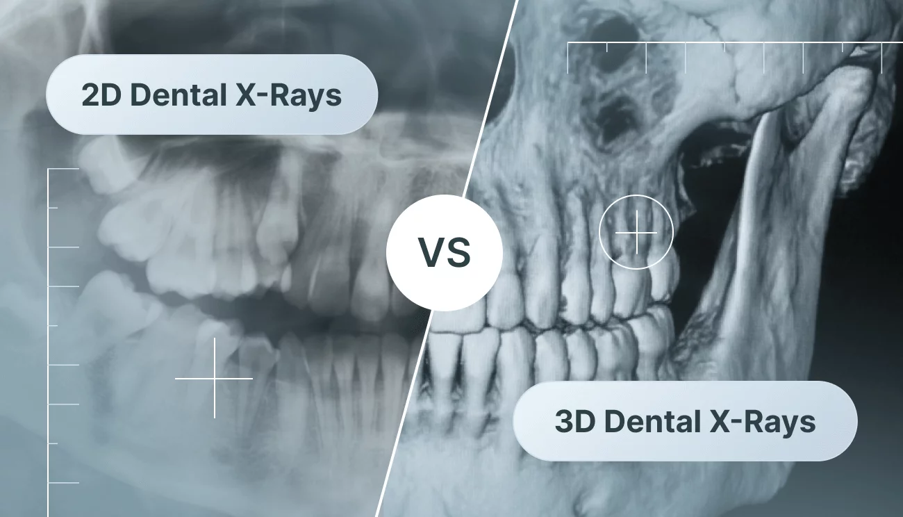
CBCT stands for cone-beam computed tomography — a specialized type of CT scan built specifically for the teeth, jaws and face. Where a regular dental x-ray gives a single flat, two-dimensional picture, a CBCT scanner makes one smooth rotation around your head and captures hundreds of images, then reconstructs them into a detailed 3D model of your anatomy. Dr. G can rotate that model on screen, slice through it at any angle, and measure structures down to the millimeter.
The payoff is a view of things flat x-rays physically can't show: the height and thickness of your jawbone, the path of the nerves running through your lower jaw, the position of your sinuses, hidden areas of infection, impacted teeth, and the true shape and number of a tooth's roots. CBCT doesn't replace your routine x-rays — it's a focused tool we reach for when seeing in three dimensions can genuinely change how we plan and protect your care.
The traditional approach vs. how we do it
Many offices still rely on flat 2D x-rays for everything — or send you across town for a 3D scan. Here's how an in-office cone-beam CT changes that.
The traditional way
- A flat 2D x-ray overlaps your anatomy onto a single plane, so bone depth, nerve position and sinus location are hidden or only estimated.
- To get a true 3D view, many offices refer you out to an outside imaging or radiology center — a second appointment, extra travel, and often a wait for the report to come back.
- Implant and surgical plans are built from flat films and clinical judgment, which can leave more room for surprises once treatment is underway.
- Findings can stay ambiguous: a flat film may hint at a problem without revealing the source of pain, a fracture, or hidden infection.
The Kentfield Dental way
- Our low-dose Vatech Green X12 captures bone, nerves, sinuses and roots in three dimensions, so depth and position can be measured precisely instead of guessed.
- The scan happens right here in Kentfield — no referral, no second trip — and the 3D model is typically ready to review together within minutes.
- That 3D data lets Dr. G map implants and extractions around your individual anatomy before starting, which is designed to reduce surprises and protect what matters.
- A clearer 3D picture can help pinpoint hidden infections, fractures and impacted teeth, turning an uncertain diagnosis into a more confident one.
Why it matters for you
Sees what flat x-rays miss
A 2D x-ray flattens everything onto one plane. CBCT reveals bone volume, nerve canals, sinus position and root anatomy in three dimensions — the details that help determine whether a treatment is safe and predictable.
Safer, more precise surgical planning
By mapping the location of nerves, sinuses and available bone before we begin, the 3D scan helps Dr. G plan implants and extractions around your individual anatomy — designed to reduce surprises during treatment.
Low radiation dose
The Green X12 is designed as a low-dose system, capturing a full 3D view with a modest exposure that's generally far less than a conventional medical CT — and we only recommend a scan when the diagnostic benefit clearly justifies it.
Done in-house, same visit
No referral to an outside imaging center and no waiting days for a report. The scan happens in our Kentfield office and we review it together within minutes, so planning can often start the same day.
Clearer, more confident diagnosis
CBCT can help uncover hidden infections, fractures, impacted teeth, cysts and the source of pain that flat films leave ambiguous — turning an uncertain picture into a clearer one.
You see exactly what we see
Dr. G rotates your 3D scan on screen and explains what it shows, so you understand the reasoning behind every recommendation and stay an informed partner in your care.
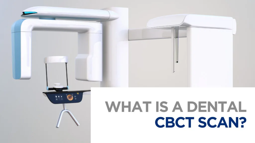
The equipment: Vatech Green X12 cone-beam CT (CBCT).
Frequently asked questions
A CBCT scan is a low-dose 3D x-ray of your teeth and jaws. Unlike a flat dental x-ray, cone-beam CT captures your anatomy in three dimensions — showing bone, nerves, sinuses and tooth roots in detail — which is why it's often the foundation for implant and surgical planning.
A regular dental x-ray is a flat, 2D image, while a CBCT scan is a 3D model we can rotate and measure. CBCT reveals bone volume, nerve position, sinuses and root anatomy that flat films overlap and hide — so it's used for implants, surgery and complex diagnosis rather than routine checkups.
Dental CBCT is generally considered safe when used appropriately. Our Vatech Green X12 is a low-dose system, and a dental cone-beam scan typically uses far less radiation than a conventional medical CT. We only recommend a scan when the diagnostic benefit clearly justifies the small exposure, and Dr. G will always explain why a scan is needed first.
In many cases, yes. A CBCT scan lets us measure your jawbone and locate the nerves and sinuses precisely, which helps us plan an implant safely and accurately. The same 3D data can also feed the surgical guide we 3D-print in-house for guided placement.
You can get it right here in Kentfield. We keep an in-office Vatech Green X12 cone-beam CT, so Marin patients don't need a referral to an outside radiology center — the scan and the review happen in our office, often in the same visit.
Most patients find a CBCT scan very easy. You simply rest your chin on a support while the scanner makes one quiet rotation around your head — nothing goes inside your mouth, there are no molds or bulky sensors, and the actual exposure lasts only seconds.